Coronary Angiography And Angioplasty
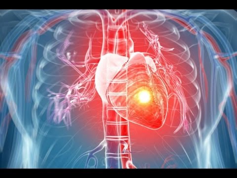
Coronary angiography is a procedure to visualize the coronary arteries. The coronary arteries are the blood vessels that supply blood to the heart muscles. To see the coronary arteries, a special dye or contrast medium is injected through a small tube (catheter) inserted via a large artery in the groin or the wrist. The catheter is then advanced to the heart and positioned at the openings of the coronary arteries before injection is performed.
Angioplasty is a procedure to expand narrow arteries that may follow on from an angiogram. Both procedures are done in hospital under local anaesthetic. Angioplasty is a treatment performed by a doctor which involves inserting a small inflatable balloon into a narrowed artery. Sometimes angioplasty may also involve putting a stent (a short tube of expandable mesh) into a narrowed artery. Angioplasty may not be suitable for everyone.
Peripheral Vascular Interventions
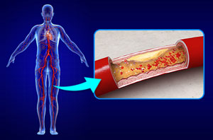
When patients suffer from hardening of the arteries, or atherosclerosis, their arteries are partially blocked by a substance called plaque. When these blockages occur in the legs or arms, they are called peripheral artery disease. Peripheral vascular interventions remove the plaque and restore the flow of blood through the artery. These interventions are medical specialties that treat peripheral artery diseases without surgically opening the leg or arm. Instead, the doctor uses small tools and at least one catheter.
A catheter is a thin tube that is inserted into a blood vessel through a small cut, usually in the leg or arm, and threaded to the site of disease. Once in place, it acts as a tunnel, enabling the doctor to efficiently guide the tools to where they are needed. By using a catheter, doctors avoid making large surgical cuts when they remove the blockage. As a result, procedures that rely on a catheter generally decrease pain, pose less risk of infection, avoid large scars and shorten recovery times. In some cases, the patient may go home the same day.
Carotid Angioplasty
Carotid angioplasty and stenting are procedures that open clogged arteries to restore blood flow to the brain. They're often performed to treat or prevent stroke. The blood vessels that bring blood to your brain and face are called the carotid arteries. The blood flow in this artery can become partly or totally blocked by fatty material called plaque. A partial blockage is called carotid artery stenosis (narrowing). A blockage in your carotid artery can reduce the blood supply to your brain. Sometimes part of a plaque can break off and block off another artery. A stroke can occur if your brain does not get enough blood.
Carotid angioplasty is often combined with another procedure called stenting. Stenting involves placing a small metal coil (stent) in the clogged artery. The stent helps prop the artery open and decreases the chance of it narrowing again.
Temporary And Permanent Pacemaker Implantation
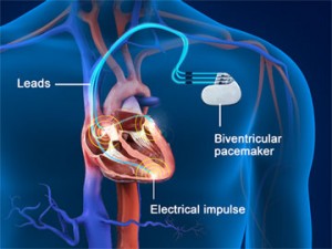
A pacemaker is an electronic device, approximately the size of a pocket watch, that senses intrinsic heart rhythms and provides electrical stimulation when indicated. There are 3 procedures which are commonly used for temporary or permamnent pacemaker implantation :
- Single-chamber pacemaker - With this device, 1 pacing lead is implanted in the right atrium or ventricle.
- Dual-chamber pacemaker - With this device, 2 pacing leads are implanted (1 in the right ventricle and 1 in the right atrium); this is the most common type of implanted pacemaker.
- Biventricular pacing (cardiac resynchronization therapy [CRT]) - With this approach, in addition to single- or dual-chamber right heart pacing leads, a lead is advanced to the coronary sinus for left ventricular epicardial pacing.
Coil Embolisation
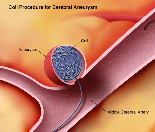
Coil embolization is a procedure to treat abnormal blood vessels in the brain and other parts of the body. It is an alternative to open surgery. This procedure cuts off the blood supply to a certain part of the body.
A thin tube, containing the coil device on a guidewire, is inserted into a large artery, usually in the groin, and passed up into the skull under X-ray control. The coil is placed inside the aneurysm and detached from the guidewire. Multiple coils can be placed the aneurysm through the same tube until the aneurysm is densely packed.
Renal angioplasty
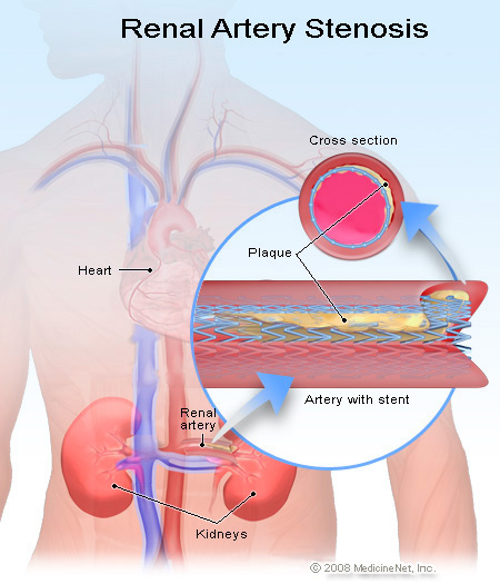
In Renal angioplasty, a small flexible tube called a catheter can be positioned inside the renal artery. An angioplasty is performed by using a catheter that has a tiny balloon attached to the end. The angioplasty balloon is inflated inside the vessel and presses any plaque (blockage) into the walls of the vessel. Renal artery stenosis a narrowing of one or both of the main arteries supplying the kidney. This causes a decrease in blood flow to 1 or both kidneys. The kidneys filter and remove waste from the blood.
Standard treatment for high blood pressure may be enough if blood pressure can be controlled and the kidneys are functioning well enough.
Device Closure For ASD, VSD, PDA

Atrial Septal Defect (ASD) : This is a congenital heart defect in which blood flows between the atria (upper chambers) of the heart. A wall called the Interatrial Septum separates the Atria. If this septum is defective or absent, then oxygen-rich blood can flow directly from the left side of the heart to mix with the oxygen-poor blood in the right side of the heart, or vice versa. This causes lower-than-normal oxygen levels in the arterial blood that supplies the brain, organs, and tissues.
Ventricular Septal Defect (VSD) : This is a defect in the ventricular septum, the wall dividing the left and right ventricles (lower chambers) of the heart. The extent of the opening may vary from pin size to complete absence of the ventricular septum, creating one common ventricle. The ventricular septum consists of an inferior muscular and superior membranous portion and is extensively innervated with conducting cardiomyocytes (cardiac muscle cells).
Patent Ductus Arteriosus (PDA) : This is a condition wherein the ductus arteriosus (fetal blood vessel) fails to close after birth. This vessel does not close and remains "patent" (open), resulting in irregular transmission of blood between the aorta and the pulmonary artery. PDA is common in newborns with persistent respiratory problems such as hypoxia and has a high occurrence in premature newborns due to underdevelopment of the heart and lungs.
Cardiac Catheterisation
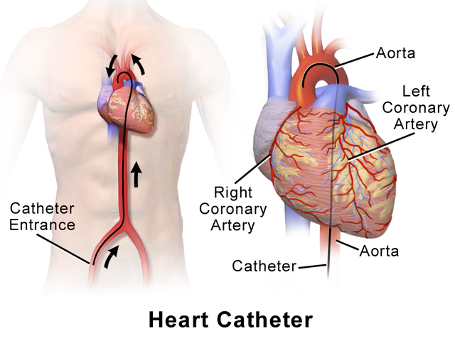
Cardiac catheterization (cardiac cath or heart cath) is a procedure to examine how well your heart is working. A thin, hollow tube called a catheter is inserted into a large blood vessel that leads to your heart. A cardiac cath provides information on how well your heart works, identifies problems and allows for procedures to open blocked arteries. Cardiac cath is usually very safe. A small number of people have minor problems. Some develop bruises where the catheter had been inserted (puncture site). The contrast dye that makes the arteries show up on X-rays causes some people to feel sick to their stomachs, get itchy or develop hives.
A doctor with special training performs the procedure with a team of nurses and technicians. The procedure is done in a hospital cardiac catheterization (cath) lab. Most people can return to their normal activities the day after the procedure depending on whether any additional interventions were done during the cardiac cath.
2D And 3D Echoca
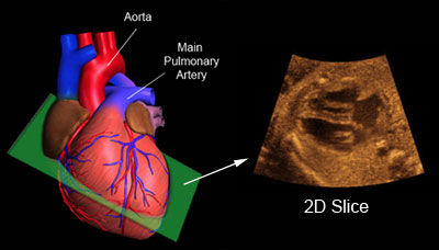
An echocardiogram is a noninvasive (the skin is not pierced) procedure used to assess the heart’s function and structures. During the procedure, a transducer (like a microphone) sends out ultrasonic sound waves at a frequency too high to be heard. When the transducer is placed on the chest at certain locations and angles, the ultrasonic sound waves move through the skin and other body tissues to the heart tissues, where the waves bounce or “echo” off of the heart structures. These sound waves are sent to a computer that can create moving images of the heart walls and valves.
Three-dimensional echocardiography (3D echocardiography) already has its established role in the evaluation of ventricular volumes and left ventricular ejection fraction (LVEF) with excellent correlation of results compared to cardiac magnetic resonance. Regarding the two-dimensional echocardiography (2D echocardiography), 3D echocardiography is closer to the real anatomy because of the absence of geometric inferences.
Preventive Cardiology - Weight loss, Cholesterol management
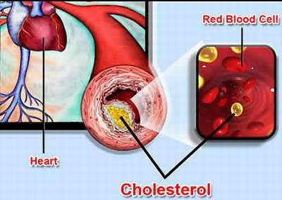
Hyperlipidemia is a medical term that means your blood has too many lipids (fats) in it. The higher your level of low-density lipoprotein (LDL) cholesterol, the "bad" cholesterol, and the lower your level of high-density lipoprotein (HDL), the "good" cholesterol, the higher your risk of cardiovascular disease. Certain genetic conditions may also cause high cholesterol levels.
Hyperlipidemia can often be improved by lifestyle changes. This is true even if high cholesterol is due to genetics. From a dietary standpoint, the best way to lower your cholesterol is reduce saturated fat and trans fat. A sedentary lifestyle lowers HDL cholesterol. Less HDL cholesterol means there’s less good cholesterol to remove LDL (bad) cholesterol from arteries. Losing excess weight can also improve cholesterol levels.